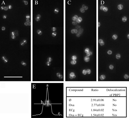
FIGURE 3.
Location of PBP2 in MRSA strain COLpPBP2-31 as determined by fluorescence microscopy. This strain carries a gfp-pbp2 fusion. The bacteria were visualized after growth in MH broth (A) and in broth containing 4 μg/ml oxacillin (B), 12.5 μg/ml ECg (C), and 4 μg/ml oxacillin and 12.5 μg/ml ECg (D). The extent of localization of PBP2 at the septum was determined by calculating the ratio (a − c/b − c), where a stands for fluorescence found at the septum, b stands for fluorescence at the lateral membrane, and c stands for background fluorescence (E). The values are the means ± S.D. from measurement of fluorescence ratios of 200 cells in each sample. A ratio (a − c/b − c) greater than 2 indicates localization at the septum, where two membranes are present, whereas a ratio of 2 or less than two indicates delocalization (59).

