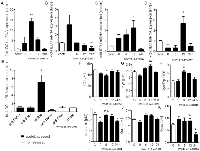
Figure 2. Transient activation of Trp catabolism after acute psychological stress.
A–E. Kinetics of IDO1 mRNA expression induced in brain (A), lung (B), spleen (C) and Peyer's patches (D) within 24-h after termination of 2-h-stress exposure (n = 6 mice/time, n = 6 controls; average values of basal expression levels in non-stressed mice were assigned as value of 1), and IDO1 mRNA expression in the brain of TNFα antiserum- and IFNγ antiserum treated vs. vehicle-treated animals 6-h after acute stress (E, n = 6 mice/group, average values of basal expression levels in vehicle treated, non-stressed mice assigned as value of 1). F–I. Kinetics of plasma concentrations of Trp (F), Kyn (G), the Kyn/Trp ratio (H) and of the levels of serotonin (I), Quin (J) and Kyna (K) following 2-h-stress exposure (n = 6 mice/time) compared with basal levels of healthy controls (n = 15 mice/group). All panels depict data of one representative experiment of two: *p<.05; **p<.01; ***p<.001 compared with non-stressed controls; #p<.05; ##p<.01; ###p<.001 compared with mice immediately after acute stress (0-h) and ⊥<.05 ⊥⊥<0.01, ⊥⊥⊥<0.001 compared with mice 24-h after stress exposure by Kruskal-Wallis testing with post-hoc Dunn Multiple comparison testing (KW- and p-values are indicated in the graph).

