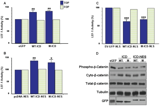
Figure 3. LRP6-ICD differentially impacts TCF/LEF-1 activity in a spatially relevant manner.
HEK 293T cells were transfected with luciferase assay reporter constructs along with (A) control eGFP, wild type GFP-LRP6-ICD (WT-ICD) or GFP-LRP6-ICD×5 m (M-ICD) for 24 hrs (B) control pcDNA-NES, wild type GFP-LRP6-ICD-NES (WT-ICD-NES or GFP-LRP6-ICD×5 m-NES (M-ICD-NES) for 48 hrs (C) control EV-GFP-NLS, wild type GFP-LRP6-ICD-NLS (WT-ICD-NLS) or GFP-LRP6-ICD×5 m-NLS (M-ICD-NLS) for 24 hrs and TCF/LEF-1 activity was measured. TCF/LEF-1 activity for the LRP6-ICD constructs is expressed as a percentage of the relevant control used in each condition: (*P<0.01, **P<0.001, ***P<0.0001). (D) HEK 293T cells were transfected with control eGFP, WT-ICD, M-ICD, WT-ICD-NES or M-ICD-NES. Twenty-four hrs post-transfection, cells were collected and cytosolic fractions were probed for endogenous cytosolic phospho-β-catenin (Ser33/37/Thr41) or total endogenous cytosolic β-catenin. Total cellular β-catenin levels did not change.

