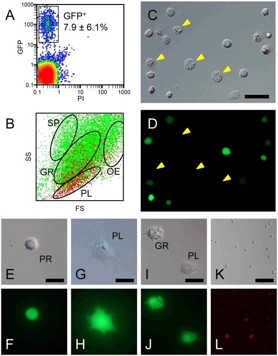
Figure 1. Ex vivo differentiation from progenitor cells in HPO.
CecB-GFP HPOs were enzymatically dispersed and injected into the hemocoel of non-transgenic larvae. Five days later, collected hemocytes were analyzed (A, B) and GFP-expressing cells were sorted (C–L). A: two-dimensional plots with PI/GFP of whole collected hemocytes. B: two-dimensional plots with FS/SS of whole collected hemocytes. Green and red dots are of GFP negative and positive cells, respectively. Clusters of spherulocytes (SP), granulocytes (GR), plasmatocytes (PL), and oenocytoids (OE) are gated according to Nakahara et al. (2009). C, D: All sorted cells fluoresce bright green, except for oenocytoids (yellow arrowheads) that had collapsed immediately after sorting. Among the GFP-expressing cells, prohemocytes (E, F), plasmatocytes (G, H), and granulocytes (I, J) were also observed. The sorted GFP+ cells were stained with anti-granulocyte antibody (K, L). Bar = 20 µm (C), 10 µm (E, G, I), 40 µm (K).

