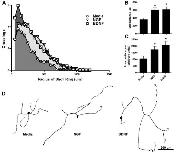
Figure 4. Semi-automated Sholl analysis detects increased axon growth in response to the growth factors NGF and BDNF.
Quantification of neurite outgrowth from DRG neurons treated with media alone (control) or media supplemented with NGF or BDNF (n=21-25/group). A) Sholl profiles generated with the semi-automated analysis revealed increased numbers of axon crossings (indicative of increased axon branching and length) after NGF or BDNF treatment compared to control. B) The maximal distance of axon growth away from the soma was significantly increased with NGF and BDNF treatment compared to control. C) The area under curve produced in (A) was significantly larger with NGF or BDNF treatment vs. control. D) β-tubulin III-stained cellular profiles representative of the statistical average for axon length and total crossings from cultures treated for 24hrs with media, NGF, or BDNF. *p<0.05 vs. media.

