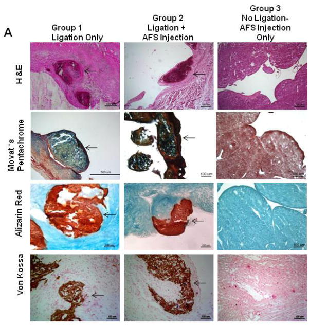
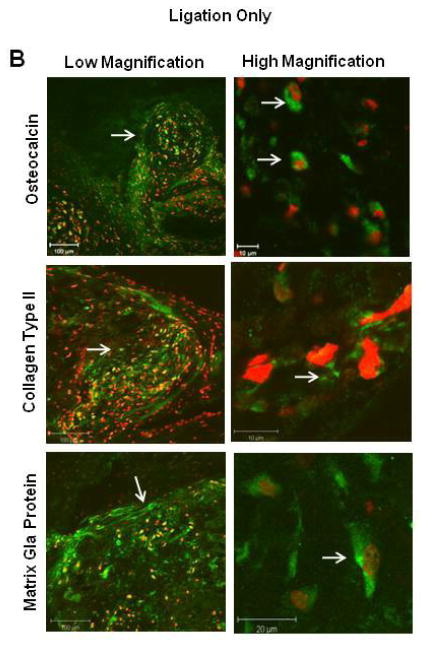
Figure 2. Characterization of Chondro-Osteogenic Masses.
(A) Representative histological examination after 3 months of Group 1 (ligation only) vs. Group 2 (ligation plus AFS injection). Both groups revealed similar chondro-osteogenic masses in the subendocardial border of the infarcted heart. Hematoxylin and Eosin staining demonstrated that the cells were arranged in a random manner with osteogenic and chondrogenic cell morphologies The masses expressed bone (yellow), collagens (blue), and mucins (red) demonstrated by Movat’s Pentachrome stain. Alizarin Red and Von Kossa assays confirmed production of calcium. (B) Immunohistochemical analysis of representative sections from Group 1 of the chondro-osteogenic masses formed in the infarcted rat hearts revealed osteocalcin, collagen type II, and Matrix-Gla-Protein staining.


