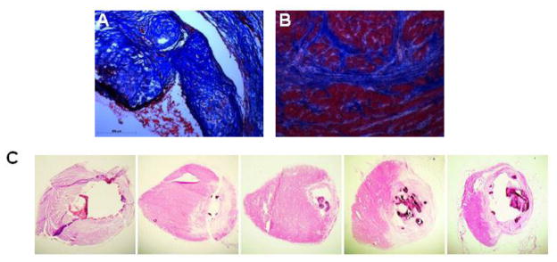
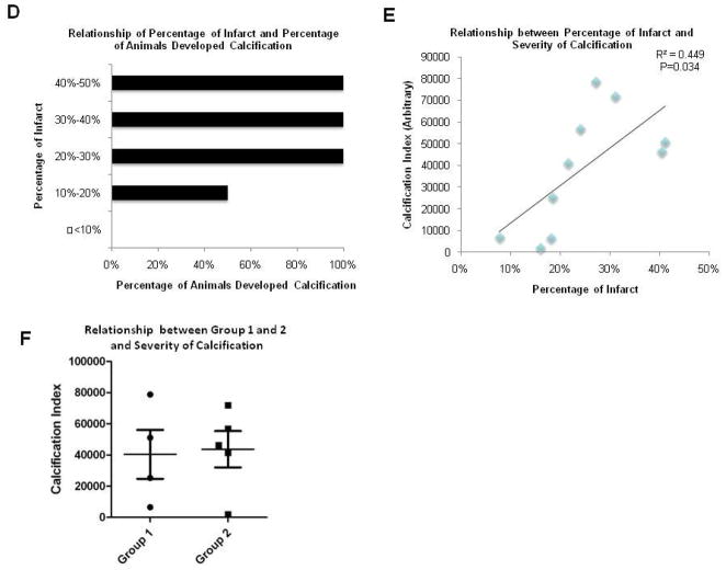
Figure 4. The Formation of Chondro-Osteogenic Masses are Related to Increased Infarction Size.
A,B. Trichrome stainings of the calcification area and the infarcted border, respectively. C. Representative serial sections of a heart stained by H&E displaying chondro-osteogenic masses. D. Comparison of infarction sizes of rats that showed chondro-osteogenic masses to rats with no masses showed a significant difference between the two groups. As the infarction percentage increased, the percentage of animals that developed calcifications also increased. E. Calcification index as a function of the infarction percentage. There is a significant linear relationship between the calcified index and the infarction percentage (r2=0.4134 and p value=0.034). F. Calcification indexes of Group 1-ligation only and Group 2-ligation plus injection of hAFS cells suggest that there is no significant difference between the calcification index of the two groups (p-value= 0.86).


