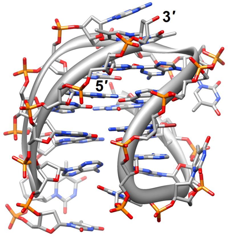
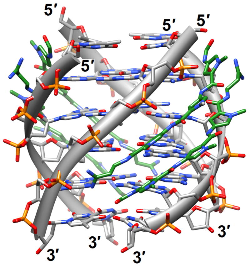
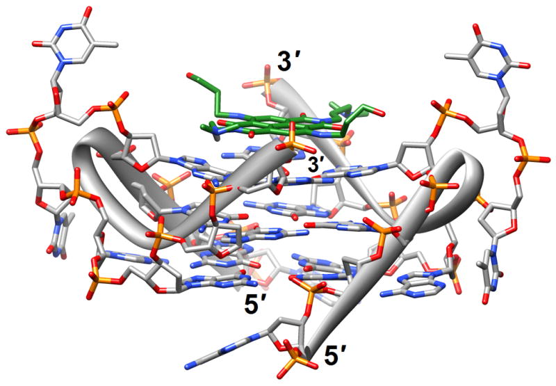
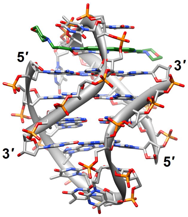
Figure 1.


G-quadruplex topology and recognition. (a) The potassium-form structure of d(G3T2A)3G3T [19] (2kf7). (b) Groove interactions between distamycin A and the parallel-stranded quadruplex [d(TG4T)]4 [22] (2jt7). (c) Stacking of tetra-substituted naphthalene diimides on the terminal G-tetrads of the parallel-stranded quadruplex [d(TAG3T2AG3T)]2 [24] (3cco). (d) Acridine ligand interactions with the antiparallel-stranded G-quadruplex [d(G4T4G4)]2 [26] (3em2). DNA atoms are colored silver, red, blue and orange for carbon, oxygen, nitrogen and phosphorus, respectively, and carbon atoms of drug molecules are green.
