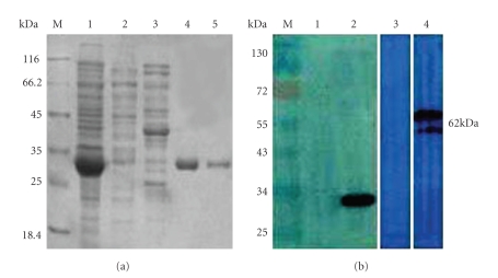
Figure 3.
SDS-PAGE and Western blot analyses of rCYP4G19. (a) M, molecular weight markers; lane 1, cell extract from pET-28a-rCYP4G19-transformed E.coli; Lane 2,3, cell extract from pET-28a- transformed E.coli; Lane 4,5, purified rCYP4G19 protein. (b) Immunoblot analysis. IgG antibodies specific to rCYP4G19 and native CYP4G19 were detected on immunoblotting membrane using sera of immunized mice (lane 2,4). Adjuvant-treated control mouse serum (negative control) is shown in lane 1,3.

