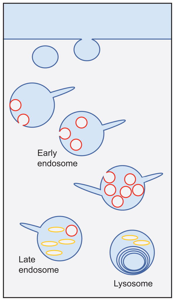
Figure 4.
MVB anatomy. Schematic of the endocytic pathway indicating the progression from early endosomes to multivesicular late endosomes and finally to multilamellar lysosomes. Internal vesicle formation occurs during endosome maturation. Pictured are two types of internal membranes: phosphatidylinositol 3-phosphate [PI(3)P] positive (red ) and LBPA positive ( yellow).

