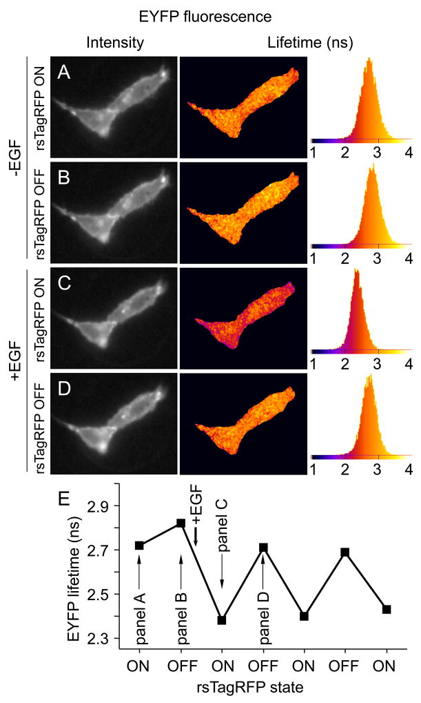
Figure 5.
FLIM-based pcFRET of EGFR-EYFP and Grb2-rsTagRFP chimeras co-expressed in live HeLa cells. For FLIM measurements EGFR-EYFP was excited at 514 nm. First, the EYFP lifetime was measured in the absence of EGF, with rsTagRFP switched ON after a 2 s exposure using 470 nm light (panel A). Subsequently, the EYFP lifetime was measured still in the absence of EGF, but with rsTagRFP switched OFF after a 2 s exposure with 470 nm light (panel B). After addition of EGF, a substantial decrease in the EYFP fluorescence lifetime due to FRET could be detected with rsTagRFP switched ON (C), and the lifetime decrease was reversed after switching rsTagRFP OFF with 577 nm (D). ON-OFF switching was repeated in the presence of EGF. In AD from left to right the DC intensity image, the pseudocolored EYFP phase lifetime-image and lifetime-histogram with ns time scale are depicted. In E panel, the average over a whole cell EYFP phase lifetime is plotted for the seven FLIM experiments with approximately 2 min steps between the subsequent FLIM measurements. See also Movie S2.

