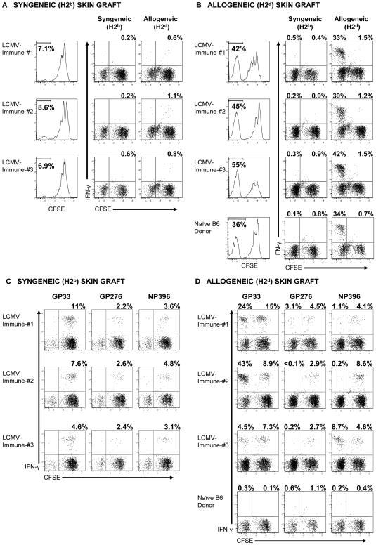
Figure 3. H2d-skin allografts stimulate LCMV-specific memory CD8 T cells to proliferate in vivo.
CFSE-labeled splenocytes (3 × 10⁷) from LCMV-immune B6 mice were adoptively transferred into naïve congenic recipient mice, which were engrafted with either syngeneic (B6, H2b) skin (A and C) or BALB/c (H2d) skin (B and D) 1 day later, as described in the Materials and Methods. (A and B) Thirteen days after engraftment, donor CD8 T cells (CD45.2) from the spleens of recipient mice were evaluated for division by dilution of CFSE, and the values represent the percentage of CD8 T cells that are CFSElo. Recovered splenocytes were incubated with either syngeneic (H2b) or allogeneic (H2d) splenocytes for 5 hr and then evaluated for the production of IFN–γ. (C and D) Alternatively, recovered splenocytes were incubated with the indicated peptides for 5 hr and then evaluated for the production of IFN–γ. For analysis, samples were gated on CD8+ cells, and the values shown represent the percentage of either CFSElo or CFSEhi CD8 T cells staining positive for IFN–γ. The data are representative of 4 experiments.

