
FIGURE 3.
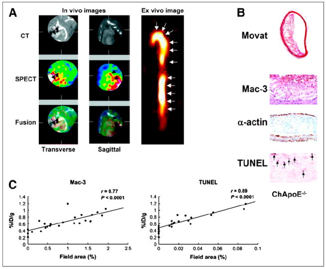
(A) Six reconstructed slices from in vivo hybrid small-animal SPECT/CT scan after injection of 99mTc annexin AV into 62-wk apoE−/− mouse fed high-fat diet and showing uptake of tracer in aortic arch (red arrows). Image on right shows excised aorta imaged ex vivo. (B) Immunohistochemical stained sections through aorta shows American Heart Association class IV lesion with lipid core, prevalent macrophages, and TUNEL-positive nuclei. (C) Correlations between percentage injected dose (%ID) and both macrophages and TUNEL-positive cells. (Reprinted from (35).)