
FIGURE 6.
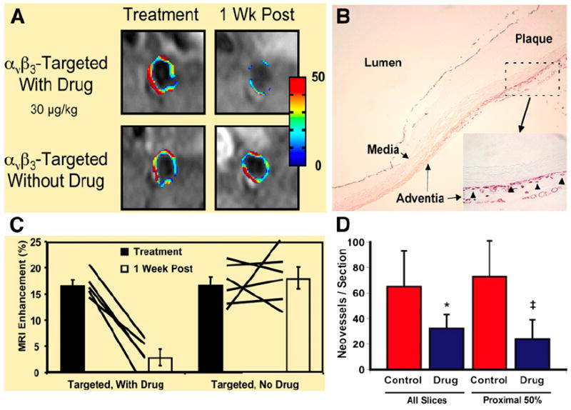
(A) MRI of abdominal aorta shows false-colored overlay of percentage signal enhancement at time of treatment (left) and 1 wk after treatment (right). (B) Platelet endothelial cell adhesion molecule (PECAM)–stained section (×4) of abdominal aorta from hyperlipidemic rabbit shows adventitia, media, and plaque. Higher-magnification inset (×20) shows that microvessels were predominantly in adventitia associated with thickening neointima. Neovessels were generally not in regions where plaque progression was minimal or nonexistent in this cohort of rabbits. Arrowheads illustrate type of PECAM microvessels counted within each section to assess fumagillin antiangiogenic effects. Larger, mature vessels positively staining for PECAM were not included in these estimates. (C) Graph of aortic MRI signal enhancement averaged over all imaged slices at time of treatment (black bars) and 1 wk after treatment (white bars). Solid lines indicate individual animal’s response to treatment over 7-d period. (D) Graph showing that number of neovascular vessels within adventitia was reduced (*P < 0.06; ‡P < 0.05) in fumagillin-treated rabbits over proximal half of aorta (i.e., renal artery to diaphragm), which correlated with region of greatest MRI signal and fumagillin response in imaging studies. (Adapted with permission of (82).)