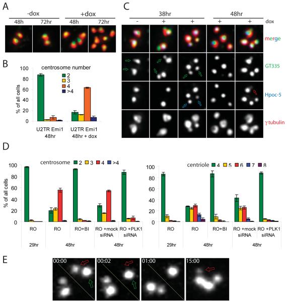
Figure 4.
Activated Plk1 is necessary for centriole reduplication. (A-C) Expression of phosphomimicking Plk1 mutant (T210D) induces centriole reduplication in Emi1-depleted U2TR cells. Mitotic cells (collected by shake-off) were plated on coverslips and 1.5 hr later transected with Emi1 siRNA. Doxycycline (1 μM) was added 24-26h after shake off. (A) Typical centrosome configurations in non-induced (−dox) vs. induced (+dox) Emi1-depleted U2TR cells. Red, γ-tubulin (PCM); green, polyglutamylated tubulin (centrioles). (B) Percentage of cells with various numbers of centrosomes under two treatment conditions. (C) Procentrioles mature and disengage from mother centrioles upon expression of Plk1(T210D). Notice that in non-induced cells all centrioles are duplicated (green arrows in 38 hr −dox). However, procentrioles are small and do not contain hPOC-5 which is normally recruited to the distal end of growing procentrioles during G2. In induced cells, procentrioles become more prominent (green arrows in 38 hr +dox) and eventually do recruit hPOC-5 (blue arrows in 38 hr +dox). At a later time, induced cells contain both individual centrioles and diplosomes. hPOC5 is found in all individual centrioles. However, some procentrioles lack hPOC-5 or contain minimal amounts of this protein (red arrow, 48 hr +dox). (See also Figure S4). (D-E) Inhibition of Plk1 prevents centriole reduplication but not initiation of procentriole assembly. Mitotic HeLa cells (collected by shake-off) were plated on coverslips and 1.5 hr later treated with RO. 100 nM Plk1 inhibitor BI2536 was added 29 hr after shake-off. Alternatively, 1.5 hr after shakeof cell were transfected with siRNA against Plk1. (D) Percentage of cells with various centriole and centrosome numbers under different conditions. (E) Procentriole assembly in G2-arrested HeLa cells after selective ablation of the original procentriole. A procentriole (red arrow in 00:00) was ablated within the diplosome (compare 00:00 and 00:02) at 31h after shake-off. This operation resulted in the formation of a new procentriole (arrow in 15:00) on the mother centriole, as evidenced by centrin-GFP signal. (See also Figure S5).

