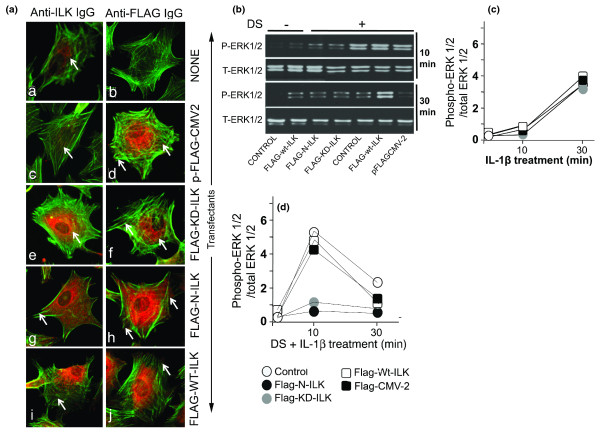
Figure 3.
Mechanical signals activate integrin-linked kinase (ILK). (a) Articular chondrocytes (ACs) either were not transfected [a,b] or were transfected with p-FLAG-CMV2 empty [c,d], FLAG-KD-ILK [e,f], FLAG-N-ILK [g,h], or FLAG-WT ILK [i,j]. ACs were immunostained with anti-ILK (left frames) or anti-FLAG (right frames) antibodies and CY3-conjugated secondary antibodies. All cells were counterstained with fluorescein isothiocyanate-phalloidin to visualize β-actin. Western blot analysis shows ERK1/2 activation in untransfected ACs or those transfected with FLAG-N-ILK, FLAG-KD-ILK, FLAG-WT-ILK, or pFLAG-CMV2 exposed to (b) no strain or dynamic strain (DS) alone, (c) interleukin-1-beta (IL-1β) alone, or (d) DS and IL-1β. Frames (c,d) show semiquantitative estimation of bands in Western blots. All figures represent one of three similar experiments. ERK1/2, extracellular receptor kinase 1/2; FLAG, polypeptide protein sequence DYKDDDDK; KD, kinase-deficient; P-ERK1/2, phospho-Thr202/Tyr204-extracellular receptor kinase 1/2; T-ERK1/2, total extracellular receptor kinase 1/2; WT, wild-type.

