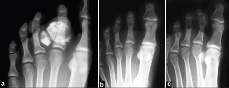
Figure 2.

(a) Antero-posterior radiograph of the right foot showing two calcified lesions in relation to the proximal phalanx of the 2nd toe. Radiograph of the right foot (b) anteroposterior view, and (c) oblique view, showing no recurrence at four years follow-up
