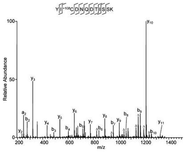
Figure 3.
An in vitro-modified peptide with a mass shift of +108 at cysteine. BSA (300 ng) was resolved by SDS-PAGE and stained with colloidal Coomassie staining solution composed of 9 vol of G−250 stain solution (ProtoBlue, National Diagnostics, Atlanta, GA) and 1 vol of ethanol. The gel band was destained with buffer composed of ethanol/water (50:50, v/v) before in-gel digestion and HPLC/MS/MS analysis.

