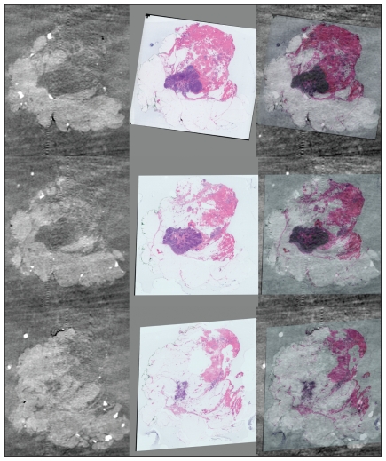
Fig. 2.
Correlation between imaging histology and 3-dimensional (3-D) reconstruction for comprehensive margin assessment. Whole-mount sections in series are digitally scanned. Each whole-mount section may be compared with slices imaged by micro-computed tomography (CT). The CT and digital images are then coregistered to allow 3-D reconstruction. Photograph courtesy of Dr. Gina Clarke.

