Figure 7.
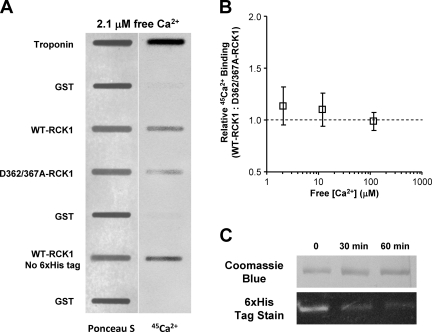
45Ca2+ binds to both WT and D362/367A-RCK1 domains. (A) Protein (Ponceau S) staining blotted on nitrocellulose membrane (left) and 45Ca2+ overlay phosphor image of the same blot (right) in 2.1 µM of free [Ca2+]. Signal intensity is proportional to protein amount (left) and Ca2+ binding (right). (B) The ratio of WT to D362/367A-RCK1 Ca2+ binding (normalized by protein amount) is plotted versus free [Ca2+]. (C) Enzymatic removal of the 6xHis from the WT-RCK1 domain. The purified 6xHis-WT-RCK1 is incubated with 5 U/ml DAPase, and then separated on a 12.5% SDS-PAGE and stained with either Coomassie Brilliant Blue (top strip) or InVision His tag in-gel stain (Invitrogen) (bottom strip). Troponin and GST are positive and negative controls, respectively. Note that the removal of the histidine tag did not reduce Ca2+ binding.

