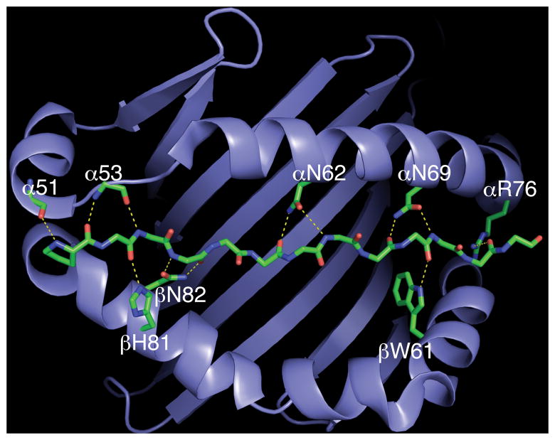
Figure 1.
The conserved hydrogen bond network in class II MHC-peptide complexes. The binding site of DR1 is represented as a ribbon diagram and the backbone of bound peptide is shown as a stick representation. Hydrogen bonds with main chain atoms of bound peptide are shown as yellow dashed lines. Main chain atoms of amino acids at DR1 positions α51 and α53 mediate hydrogen bonds, whereas conserved side chains form hydrogen bonds at other positions in the MHC molecule. The figure was generated with PyMOL software (http://www.pymol.org; PyMOL Molecular Graphics System; DeLano Scientific, San Carlos, CA) using Brookhaven Protein Data Bank coordinate file 1DLH (19).

