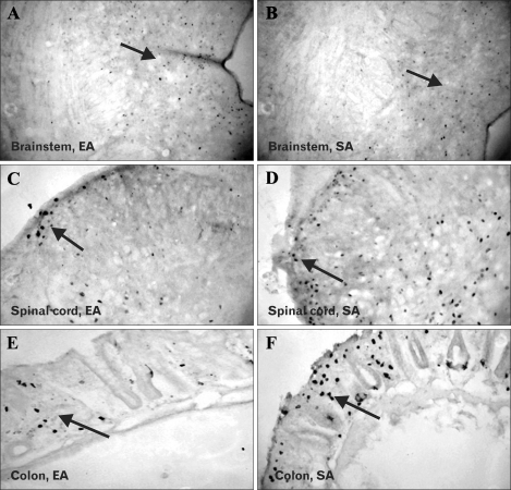
Figure 3.
Comparison of Fos expression between electroacupuncture (EA) and sham acupuncture (SA) groups. Immunohistochemistry showed significantly lower Fos expression (arrowed) in the dorsal raphe nucleus of the brain (A, EA; B, SA; ×40), laminae I and II of superficial dorsal horn of the spinal cord (C, EA; D, SA; ×40) and colonic mucosa (E, EA; F, SA; ×40) in rats treated with EA compared to those treated with SA. Fos positive cells were highly abundant on colonic epithelium among SA rats compared to EA rats.

