
Figure 1.
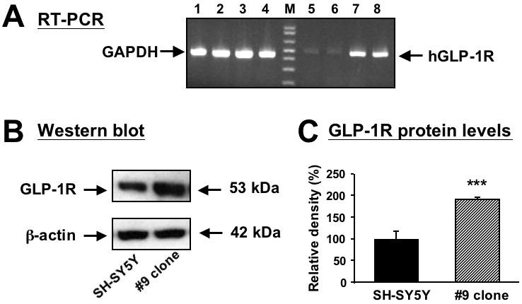
Over-expression of human GLP-1R RNA and protein in stably transfected SH-SY5Y cells. (A) RT-PCR showing human GLP- 1R mRNA expression (lanes 5 to 8) in original and human GLP-1 stably transfected SH-SY5Y (SH-hGLP-1R#9) cells. The expected RT-PCR product size is 480 bp. Glyceraldehyde 3-phosphate dehydrogenase (GAPDH) (lanes 1-4) was utilized as an external control and showed equal expression across lanes. Lanes 1, 2, 5 and 6 are non-transfected (original) cells and lanes 3,4,7 are 8 are SH-hGLP-1R#9 cells; M is a molecular ladder. (B) A representative Western blot analysis of human GLP-1R protein levels (∼53 kD) shows that hGLP-1R protein is over-expressed in SH-hGLP-1R#9 clones when compared with original SH-SY5Y cells; (C) Quantitative analysis of Western blots shows that protein levels of hGLP-1R are increased 2 fold in #9 clone as compared to original SH-SY5Y cells (normalized by β-actin levels), which was significantly different (***p<0.001, Student's t-test).