
Figure 3.
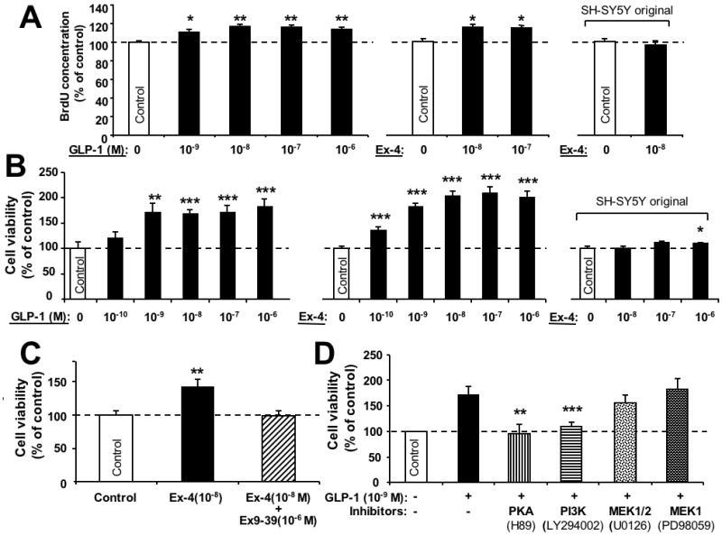
GLP-1R agonists promote cell proliferation in human neuroblastoma SH-hGLP-1R#9 cells. (A) Cell proliferation (BrdU): Increasing concentrations of GLP-1(10-9, 10-8,10-7,10-6 M) and Ex-4 (10-8, 10-7) elevated cellular BrdU concentrations after 24 h treatment versus respective controls. By contrast, Ex-4 (10-8 M) was without effect on BrdU in the original SH-SY5Y cells (right-hand side). (B) Similarly, increasing concentrations of GLP-1(10-10, 10-9, 10-8,10-7,10-6 M) and Ex-4 (10-10, 10-9, 10-8,10-7,10-6 M) increased cell viability after 24 h treatment (MTS assay). Such Ex-4 concentrations were without significant effect in the original SH-SY5Y cells (right-hand side) except at the highest concentration of 10-6 M. (C) Stimulation of cell viability by Ex-4 was abolished by simultaneous treatment of cells with the selective GLP-1R antagonist, Ex-9-39. Specifically, cells were treated with vehicle, Ex-4 (10-8M) and Ex-4 (10-8M) + Ex-9-39 (10-6 M) for 24 h, and cell viability was then determined by MTS assay. (D) Inhibition of GLP-1 (1 nM)-induced cell proliferation by specific pathway inhibitors. SH-hGLP-1R#9 cells were incubated with 10 μM H89 (PKA), 10 μM LY294002 (PI3K), 5 μM U0126 (MEK1/2) or 20 μM PD98059 (MEK1) for 20 min prior to GLP-1 treatment. After 24 h incubation with GLP-1, cell viability was evaluated by MTS assay and results are presented as a percentage of controls, n=6. All data are presented as mean ± S.E.M. Statistical evaluation, (A,B,C,D): Dunnett's t-test, p= *<0.05, **<0.01. ***<0.001, versus respective control.