
Figure 4.
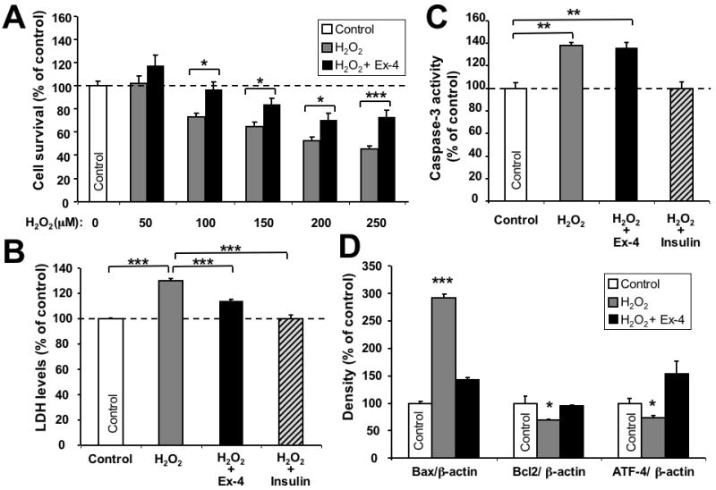
Neuroprotective effect of GLP-1 against H2O2-induced oxidative stress. (A) Ex-4 protected SH-hGLP-1R#9 cells from a wide concentration range (50, 100, 150, 200 and 250 μM) of H2O2-induced cell death. Cells were pretreated with 10-6 M Ex-4 for 2 h and, thereafter, exposed to different concentrations of H2O2 for 24 h. 100 μM H2O2 and greater concentrations induced significant cell death (Dunnett's t-test vs. no H2O2, p<0.05), and Ex-4 ameliorated H2O2-induced effects; (B) Ex-4 (10-7 M) pretreatment prevented H2O2 (100 μM) -induced elevation in LDH levels; insulin (10-8 M) was used as a positive control. (C) Caspase-3 activity assay: Ex-4 (10-7 M) pretreatment for 2 h proved unable to reverse H2O2-induced caspase-3 activity, whereas insulin pretreatment was capable of doing so. (D) Western blot analysis of Bax, Bcl-2 and ATF-4 protein levels. β-actin was used as an internal standard to normalize protein levels. Data are presented as mean ± S.E.M. and as a percentage of controls. Statistical evaluation, (A): unpaired student's t-test and one-way ANOVA; (B,C,D): Dunnett's t-test, p= *<0.05, **<0.01. ***<0.001, N=6.