
Figure 5.
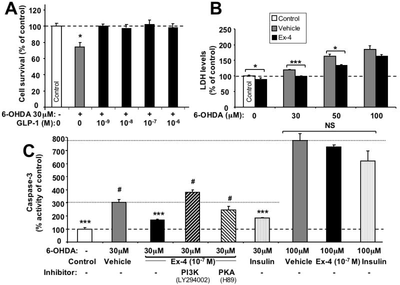
Neuroprotective effect of GLP-1 against 6-hydroxydopamine- (6-OHDA)-induced cell death. (A) Increasing concentrations (10-9, 10-8,10-7,10-6 M) of GLP-1 pretreatment for 2 h protected SH-hGLP-1R#9 cells from 30 μM 6-OHDA induced cell death: MTS assay at 24 h after 6-OHDA exposure. (B) Ex-4 (10-7 M) pretreatment for 2 h prevented 6-OHDA (30 and 50 μM, but not 100 μM) induced elevations in LDH levels in SH-hGLP-1R#9cells. (C) Caspase-3 activity assay: SH-hGLP-1R#9 cells exposed to 30 μM 6-OHDA induced a 3.2-fold elevation in caspase-3 activity. Ex-4 (10-7 M) pretreatment for 2 h significantly ameliorated 6-OHDA induced caspase-3 activity, and insulin (10-8 M), used as a positive control, showed a similar effect. However, following 20 min preincubation of cells with a PKA (10 μM H89) or PI3K inhibitor (10 μM LY294002) prior to Ex-4 addition, the effect of Ex-4 on caspase-3 activity was largely (in the case of H89) or completely (in case the of LY294002) abolished. A 100 μM 6-OHDA insult significantly elevated caspase-3 levels by 7.7-fold and, in accord with B, neither Ex-4 (10-7 M) nor insulin (10-8 M) were able to ameliorate this action. Data are presented as mean ± S.E.M. and as a percentage of controls. Statistical evaluation, (A): unpaired student's t-test and one-way ANOVA; (B,C,D): Dunnett's t-test, p= *<0.05, **<0.01. ***<0.001, # or NS: not significantly different from one another (p>0.05), N=6.