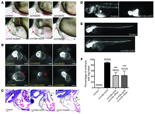
Figure 2. Expression of ccm3 proteins lacking the 18 amino acids encoded by exon 3 confers cardiovascular phenotypes characteristic of heg, ccm1, and ccm2 deficiency.
(A) Light images of the hearts of 48-hpf zebrafish control embryos, ccm2 mutant embryos, and embryos injected with morpholinos that block splicing into exon 3 of ccm3a only (ccm3aX3, 3 ng/embryo), ccm3b only (ccm3bX3, 3 ng/embryo), both ccm3a and ccm3b [ccm3(a+b)X3], or exon 2 of ccm3a (ccm3aX2, 3 ng/embryo) are shown. Arrows indicate the embryo hearts. (B) Fluorescence images of the hearts of transgenic embryos in which myocardial cells express GFP following injection of the indicated morpholinos. ccm2MO indicates a morpholino that blocks splicing of the ccm2 gene. (C) Thinned myocardium in ccm3(a+b)X3 morphants is identical to that seen in ccm2 mutants. Shown are hematoxylin/eosin-stained sagittal sections of the indicated 48-hpf embryos. a, atrium; v, ventricle; hw, heart wall. (D) Angiography of 48-hpf control and ccm3(a+b)X3 morphant embryos reveals blocked circulation at the cardiac outflow tract. (E) Vascular endothelial patterning as revealed in Tg (fli1a:EGFP)y1 embryos is undisturbed in ccm3(a+b)X3 morphant embryos. The images are composites of 2–3 images taken of the same embryos. (F) The big heart phenotype conferred by morpholinos that block splicing into exon 3 of ccm3a and ccm3b is rescued by coinjection of cRNAs (100 pg/embryo) encoding either ccm3a or ccm3b (right 2 bars). Shown are mean and SEM. The number of embryos examined is indicated above each bar, and the number of injections performed for each group shown in parentheses. ***P < 0.001 by Student’s t test. Scale bars: 20 μm.

