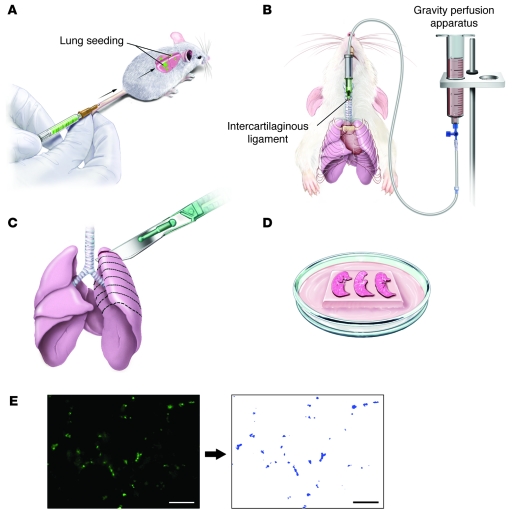
Figure 1. Schematic diagram summarizing the PuMA experimental approach.
(A) GFP-positive tumor cells (2 × 10⁵) were delivered to mice by tail-vein injection. (B) Following humane euthanasia, the trachea was cannulated with 20-gauge intravenous catheter and attached to a gravity perfusion apparatus. The lungs were infused in the vertical position under a constant 20 cm H2O hydrostatic pressure. (C) The lungs were allowed to cool at 4°C for 20 minutes to solidify the agarose medium solution. Complete transverse serial sections (1–2 mm in thickness) were gently sliced from each lobe with a scalpel, yielding 16–20 lung slices per pair of lungs. (D) 4–5 lung sections were placed on the sterile Gelfoam sections bathing in culture media. (E) Images were acquired and the area of GFP-positive cells in each lung was quantified. Scale bars: 200 μm.

