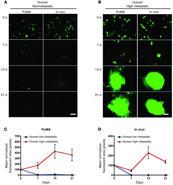
Figure 5. Similarities in metastatic progression in vivo compared with PuMA.
A direct comparison of the metastatic phenotype of human osteosarcoma cell lines in vivo (experimental metastasis) and in the PuMA was conducted. (A and B) Serial imaging of fluorescently labeled high- and low-metastatic human osteosarcoma cells was conducted in the PuMA. At the identical time points, lungs from mice that had received tail-vein injection of tumor cells were collected and imaged as in the PuMA. Patterns of pulmonary metastatic progression were similar in both in vivo and PuMA. Representative fields from lung are shown. Scale bars: 200 μm. (C and D) Quantification of metastatic burden (mean normalized fluorescent area) from A and B. Identical results demonstrating the similarities in pulmonary metastatic progression for murine osteosarcoma cells was seen in vivo and in the PuMA. Plotted data represent the mean ± SD.

