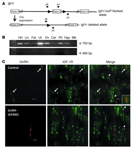
Figure 3. Generation of the GnRH-IGFRKO mouse.
(A) The Igf1r gene with exon 3 flanked by loxP sites (large arrows) before (top panel) and after (bottom panel) Cre recombination. Primers used for detection of the truncated Igf1r gene are labeled p4, p5, and p6. (B) PCR analysis of genomic DNA isolated from tissues of a GnRH-IGFRKO mouse. The floxed Igf1r gene is indicated by the 750-bp band, and the excised Igf1r gene is indicated by the 490-bp band. (C) Sections (40 μm) of hypothalami from control littermates or GnRH-IGFRKO mice were incubated with anti-GnRH and anti–IGF-1R antibody with secondary antibodies conjugated to a fluorophore emitting red (GnRH) or green (IGFR) wavelengths. Long arrows indicate neurons that stain for GnRH and IGF-1R. Short arrows indicate neurons that stain for IGF-1R only. Scale bars: 20 μm.

