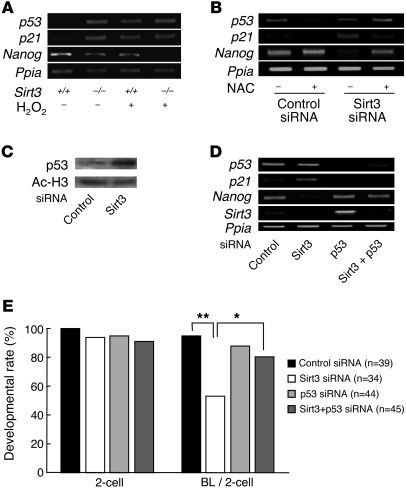
Figure 10. Involvement of p53 in developmental arrest of Sirt3-deficient preimplantation embryos.
(A) p53 and p21 were upregulated in Sirt3–/– embryos as in H2O2-treated wild-type embryos. Nanog expression was decreased in Sirt3–/– embryos, and the decrease was enhanced by H2O2 stimulus. (B) Effects of Sirt3 knockdown and treatment with NAC on the expression of p53 and its downstream genes. In Sirt3 siRNA–injected embryos, p21 expression was upregulated, whereas Nanog expression was downregulated. These effects were blocked by NAC. (C) Western blotting analysis showing increased p53 protein levels in Sirt3-knockdown embryos at the morula stage. Signals for acetylated histone H3 (Ac-H3) served as an internal control. (D) Effects of p53 knockdown on Sirt3 siRNA–induced changes in the expression of genes downstream of p53. Sirt3 siRNA–induced p21 upregulation and Nanog downregulation were blocked by siRNA-mediated p53 knockdown. Ppia expression served as an internal control in A, B, and D. (E) Effects of p53 knockdown on preimplantation developmental arrest in Sirt3-knockdown embryos. The rate of blastocyst formation was significantly improved by coinjection with p53 siRNA. Data are derived from 4 independent experiments. Statistical assessments were performed by applying Ryan’s multiple-comparison test. *P < 0.05; **P < 0.001.

