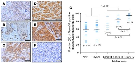
Figure 1. Elevated Sema3E expression in invasive human melanomas.
(A–F) Sema3E expression was detected by immunohistochemistry in a series of 58 nevi and melanoma human samples, representing different stages of progression. Micrographs show representative fields of Sema3E-expressing samples: (A) an intradermal nevus, (B) a dysplastic nevus, (C) a Clark III level malignant melanoma, and (D and E) 2 different Clark IV level melanomas. (F) Control staining of a Clark IV melanoma, without including the specific antibody. Scale bar: 40 μm. (G) In the graph, each case is represented by a blue bar, indicating the fraction of Sema3E-positive cells in the sample (scored as described in Methods). Black bars indicate the mean value for each of 5 groups (intradermal-junctional nevi [nevi], dysplastic nevi/melanomas in situ [dyspl.], and Clark II, Clark III, and Clark IV level melanomas).

