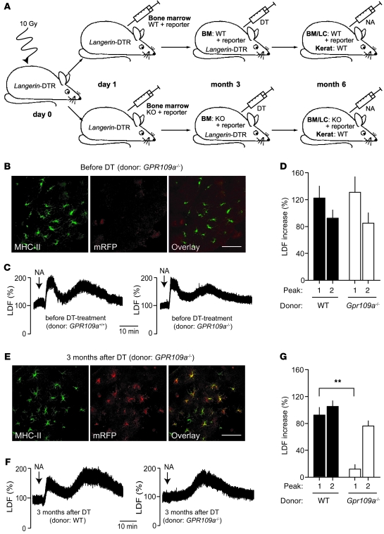
Figure 2. GPR109A expressed by Langerhans cells mediates only the early phase of nicotinic acid–induced flushing.
(A) Experimental scheme. At 1 day after irradiation, Langerin-DTR mice were transplanted with BM from WT or Gpr109a–/– mice carrying in both cases Gpr109amRFP (reporter). At 3 months after transplantation, both groups were treated with DT, and 3 months later, nicotinic acid–induced flushing was evaluated. LC, Langerhans cells; kerat, keratinocytes. (B) Fluorescence image of epidermal sheet from transplanted animals before DT treatment stained with an anti–MHC-II antibody and analyzed for mRFP expression. (C and D) Nicotinic acid (250 μg/g) induced flushing in both experimental groups before DT treatment. (E) Fluorescent images of epidermal sheets of transplanted animals stained with anti–MHC-II antibodies and analyzed for mRFP expression 3 months after DT treatment. (F and G) Effect of nicotinic acid on flushing in mice transplanted with WT and Gpr109a–/– BM 3 months after DT treatment. Scale bars: 20 μm. Data are representative of at least 3 independent experiments. Results in D and G are mean ± SEM (n ≥ 6). **P ≤ 0.01.

