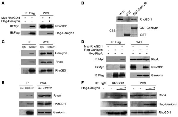
Figure 3. Gankyrin interacts with RhoGDI1 and increases the association of RhoGDI1 and RhoA.
(A) Immunoassay of HEK293 cells cotransfected with vectors expressing Flag-Gankyrin and Myc-RhoGDI1. Lysates were immunoprecipitated with anti-Flag and then analyzed by immunoblot with anti-Myc and anti-Flag antibodies. WCLs, whole cell lysates. (B) Immunoblot analysis of bound proteins in lysates of HEK293 cells expressing Myc-RhoGDI1, incubated with Sepharose beads coupled to GST alone or a fusion protein of GST and Gankyrin (GST-Gankyrin). CBB, Coomassie brilliant blue staining. (C) Immunoblot analysis of the interaction among endogenous Gankyrin, RhoA, and RhoGDI1 in lysates of HEK293 cells after immunoprecipitation with rabbit IgG or anti-RhoGDI1 (left) and whole cell lysates (right). (D) Immunoassay of HEK293 cells cotransfected with vectors expressing Flag-Gankyrin, Myc-RhoA, and Myc-RhoGDI1. Lysates were immunoprecipitated with anti-Flag and analyzed by immunoblot with anti-Myc and anti-Flag antibodies. (E) Immunoblot analysis of the interaction among endogenous Gankyrin, RhoA, and RhoGDI1 in lysates of HEK293 cells after immunoprecipitation with rabbit IgG or anti-Gankyrin (left) and whole cell lysates (right). (F) Immunoassay of HEK293 cells transfected with increasing amounts of Flag-Gankyrin. RhoGDI1 in lysates was immunoprecipitated with anti-RhoGDI1, followed by immunoblot analysis of immunoprecipitates (left) and whole cell lysates (right).

