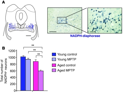
Figure 3. Loss of PPN cholinergic neurons in MPTP-treated macaques.
(A) NADPH+ neurons mapped in the PPN of a control brain of a young macaque; hatched areas denote myelinated nerve fibers in the brainstem and blue dots represent individual neurons. Low- and high-magnification images are shown at right. (B) Total number of NADPH+ neurons quantified in young and aged MPTP-intoxicated macaques compared with their respective controls (n = 4 per group). MPTP did not affect NADPH+ neurons in young animals, but induced a 30% loss of cholinergic neurons in aged monkeys. **P < 0.01, Mann-Whitney U test. Scale bars: 2 mm (map); 500 μm (low magnification); 100 μm (high magnification).

