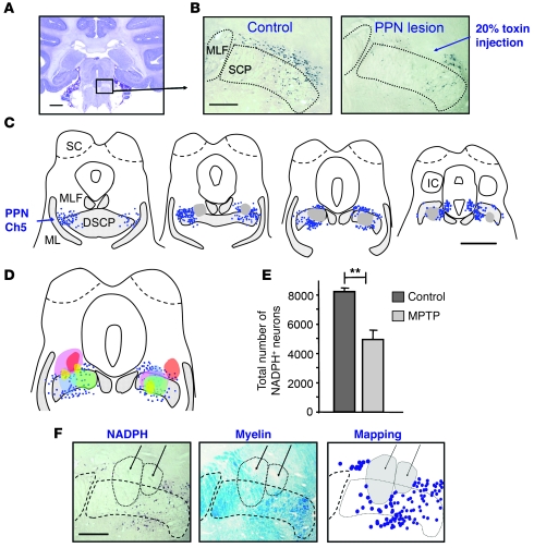
Figure 5. Loss of NADPH+ neurons after bilateral PPN lesion.
(A) Nissl-stained section showing the anatomical localization of the PPN (boxed region) in a control macaque brainstem. (B) Photomicrographs of PPN sections labeled for NADPH diaphorase histochemistry, showing a 20% toxin injection site into the PPN compared with a control. (C) Computer-generated maps of NADPH+ neurons (blue) in 4 regularly spaced sections covering the anteroposterior extent of the structure in lesioned animal M4. Each dot represents an NADPH+ neuron; gray areas represent the extent of the injection site; hatched areas denote myelinated nerve fibers. (D) All injection sites of the 5 macaques were transferred onto the corresponding brainstem map of macaque M4. Each individual is represented by a different color. Note that the cholinergic part of the PPN was lesioned in all animals. (E) Quantification of the total number of NADPH+ neurons in the PPN, showing that neuronal loss reached 39% in lesioned animals (n = 5) compared with controls (n = 5). **P < 0.01, Mann-Whitney U test. (F) Adjacent PPN sections, labeled for NADPH diaphorase histochemistry and for myelin staining with luxol fast blue, and mapping of NADPH+ neurons showed that myelinated fibers were preserved after toxin injections at 20%. Arrows indicate the center of 2 lesions. IC, inferior colliculus; SCP, superior cerebellar peduncle. Scale bars: 5 mm (A and C); 1 mm (B and F).

