
Figure 2.
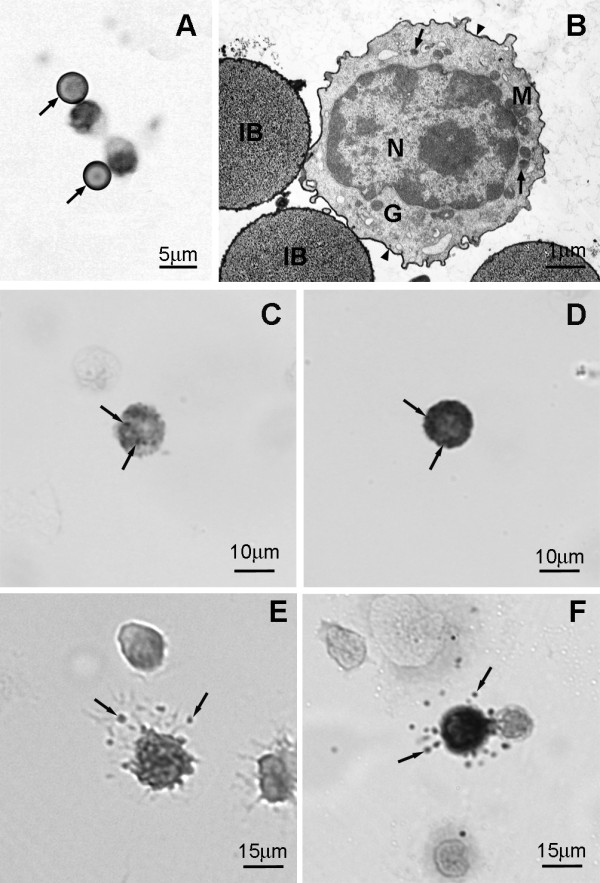
On day 2 following i.p. injection of distilled water only MCp are present in the peritoneal lavage . The MCp were isolated using magnetic beads conjugated to mAb AA4. (A) By light microscopy, the cells, which can be recognized by their attachment to immunomagnetic beads (arrows), have a large nucleus and no metachromatic granules. Toluidine blue. (B) By electron microscopy the MCp has a large centrally located nucleus (N), a few small secretory granules (arrows), and a compact Golgi complex (G). The cell surface also stains positively with anti-IgE (arrowheads). IB, immunomagnetic bead; M, mitochondria. Immature mast cells are present in the peritoneal lavage at 6 and 10 days after distilled water injection. (C) Representative immature peritoneal mast cell 6 days after i.p. injection of distilled water contains a few metachromatic granules (arrows). Toluidine blue. (D) Peritoneal mast cell, representative of the peritoneal mast cell population 10 days after i.p. injection of distilled water, contains many metachromatic granules (arrows). Toluidine blue. (E) Representative immature peritoneal mast cell 6 days after i.p. injection of distilled water is degranulated after exposure to antigen (arrows, released secretory granule matrix). Toluidine blue. (F) 10 days after i.p. injection of distilled water, this representative peritoneal mast cell, is degranulated after exposure to antigen. (arrows, released secretory granule matrix). Toluidine blue.