Figure 1.
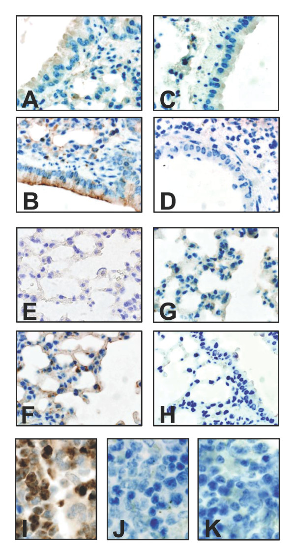
Lipocalin 2 expression in the lungs of E. coli-infected mice. Immunohistochemical staining using a polyclonal antibody against lipocalin 2 (diluted 1:250) on formalin-fixed lung sections removed 48 hours post-infection with E. coli H9049. Weak staining for lipocalin 2 is found in uninfected bronchial epithelium (A) and alveolear tissue (E) of wild-type C57BL/6 mice. Strong induction is seen following E. coli infection (4 × 10⁷ CFU E. coli H9049/mouse) in wild-type mice (B and F) whereas no staining for lipocalin 2 is seen in infected Lcn2 knock-out mice (C and G). The specificity of the reaction is demonstrated by the lack of staining when using rabbit pre-immune serum (dilution 1:250) as negative control (D and H). Staining for lipocalin 2 was also observed in neutrophils in the bone marrow of wild-type mice (I) but not in Lcn2 knock-out mice (J) or in wild-type mice incubated with pre-immune serum (K).

