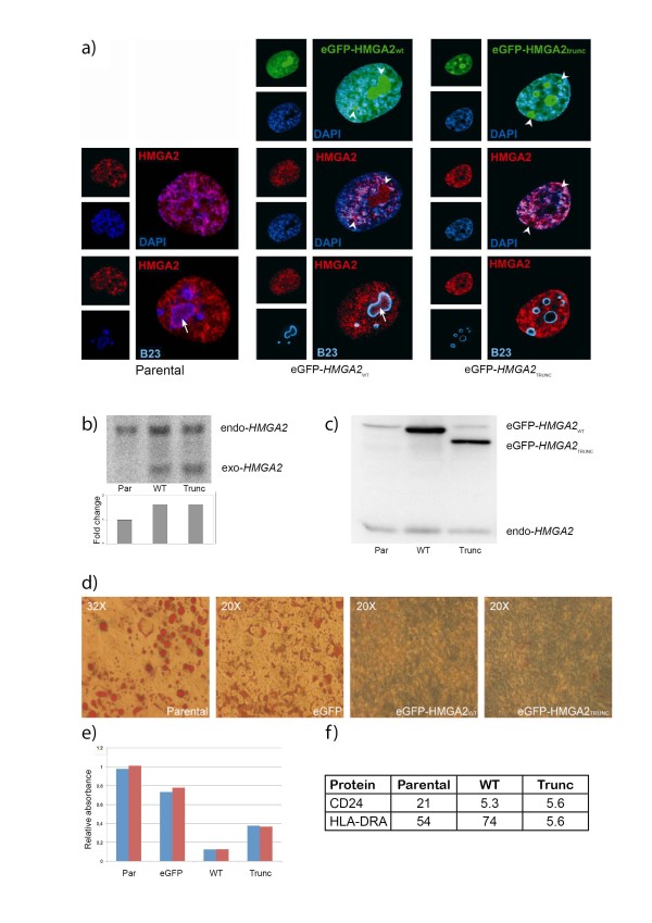
Figure 1.
Characterization of hMSC-TERT20 cells stably over-expressing eGFP-HMGA2WT and eGFP-HMGA2TRUNC. a) Nuclear localization of eGFP-tagged HMGA2 proteins was observed in hMSC-TERT20 over-expressing eGFP-HMGA2WT or eGFP-HMGA2TRUNC. Endogenous HMGA2 was visualized by immunofluorescent staining with an anti-HMGA2 antibody (The intensity in parental cells was adjusted to make observation of nucleolar fluorescence possible). The nucleolus was detected with an anti-B23 antibody, while heterochromatin was stained by DAPI, showing co-localization of HMGA2 with both nucleoli and heterochromatin. Arrowheads show concentration of endogenous or eGFP-tagged HMGA2 at sites that correspond with nucleoli, while an arrow indicates discrete foci of HMGA2 within nucleoli. b) Expression levels of endogenous and exogenous HMGA2 transcripts; expected size of the endogenous transcript is 4468 bp, while the exogenous is only 327 bp due to the lack of the 3' UTR. Top panel, northern blot of HMGA2 mRNAs. Lower panel, quantitation of total HMGA2 mRNA levels by real-time PCR, normalized for TBP expression and represented as fold induction over parental cells. c) Expression levels of endogenous and exogenous HMGA2 proteins, as detected on western blot. d) Accumulation of fat in cell cultures grown in basal medium supplemented with MDI and Rosiglitazone for 14 days. e) Relative accumulation of fat-bound Oil Red O after 14 days of differentiation of hMSC-TERT20 cells. Data from two independent experiments are shown. Par, Parental cells; eGFP, eGFP-transduced; WT, transduced with eGFP-HMGA2WT; Trunc, transduced with eGFP-HMGA2TRUNC f) Expression of CD24 and HLA-DRA on each hMSC TERT20-derived cell lines as measured by flow cytometric mean fluorescence intensity (MFI).

