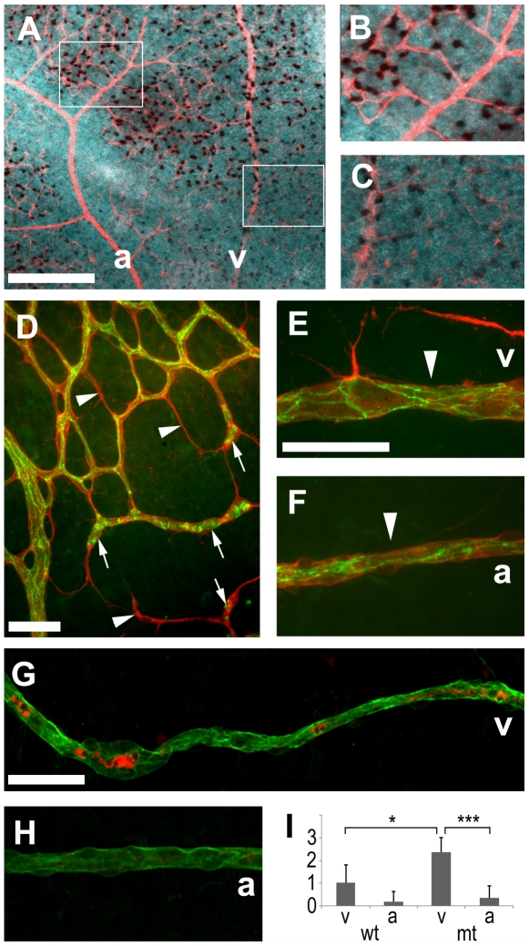
Figure 4. Vessel degeneration after one day of hyperoxia (P7–8).
(A–C) In situ hybridization showed that Vegf mRNA was detectable within the vascular network (stained with anti-collagen IV, red) but strongly reduced around arteries. Immunohistochemistry with anti-collagen IV (red D–F, green G, H), anti-claudin 5 (green D–F) and active caspase 3 (red G, H) revealed dying vessels. Empty basement membrane sleeves (arrows D) and isolated claudin 5 positive clumps were indicative of a regressing capillary network and local narrowing of artery and vein profiles (arrowheads E, F) suggested blood flow reductions. (G–I) Astrocyte-specific VEGF deletion increased radial vessel degeneration and affected veins more strongly than arteries at this early time point. Scale bars are 200 µm in A and 50 µm in D–G; * is p<0.05 and *** is p<0.001.

