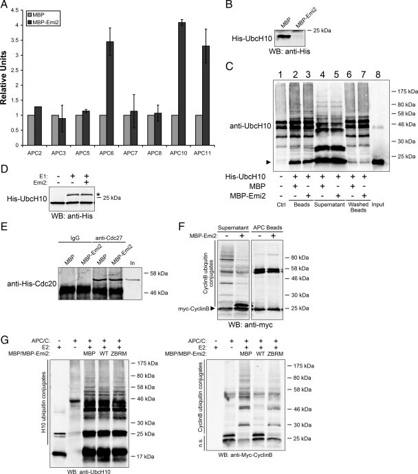
Figure 4.
Emi2 inhibits APC/C by blocking the transfer of ubiquitin from activated E2 to substrates. (A) MBP or MBP-Emi2 conjugated to amylose beads were incubated with 35S-labeled IVT APC subunits for 1 h at 22°C. Beads were washed and bound IVT APC subunits were examined by SDS-PAGE and autoradiography. Results from at least three independent experiments for each subunit were quantified with ImageQuant5.0. (B) APC/C immunoprecipitated from mitotic extracts was preincubated with MBP or MBP-Emi2 (600 nM) for 15 min at 22°C. Purified His-UbcH10 was added to both samples and incubated for 1 h at 22°C. APC/C beads were washed and the bound UbcH10 was analyzed by Western blotting. (C) APC/C was immunoprecipitated from mitotic extracts and incubated with purified His-UbcH10 in the presence of MBP or MBP-Emi2 (600 nM) for 1 h at 22°C. E1, ubiquitin, and an energy regeneration system were also added to the reactions. Beads (washed or not with PBS supplemented with 300 mM NaCl and 0.1% Triton-100) and supernatant were separated and analyzed by SDS-PAGE and immunoblotted for UbcH10. Lane 1 was APC/C immunoprecipitant without UbcH10. The arrow indicates unmodified His-UbcH10. (D) His-UbcH10 was incubated with E1, ubiquitin and an energy regenerating system in the presence or absence of 600 nM Emi2 for 30 min at 22°C. Reactions were stopped with addition of sample buffer and charging of UbcH10 was analyzed by His immunoblotting. The asterisk indicates charged/activated E2. (E) APC/C immunoprecipitated from M phase extracts was incubated in XB buffer with recombinant His-Cdc20 in the presence of 500 nM MBP or MBP-Emi2 for 20 min at 22°C. APC/C beads were retrieved and washed. The amount of associated His-Cdc20 was detected by Western blotting. (F) APC/C immunoprecipitated from mitotic extracts were incubated with precharged UbcH10, Cyclin B, and an energy-regenerating system in the presence or absence of MBP-Emi2 (600 nM) for 1 h at 22°C. Sample buffer was added to supernatant and APC/C beads separately. The formation of ubiquitin conjugates on Cyclin B was analyzed by Myc Western blotting. (G) In vitro APC/C assay was performed in the presence of 500 nM MBP or MBP-Emi2 (WT or ZBRM) and the formation of ubiquitin conjugates on both UbcH10 and Cyclin B were analyzed by Western blotting for UbcH10 or Myc. n.s., nonmodified substrates.

