Figure 5.
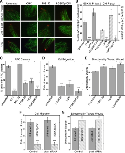
Pharmacological inhibition of GSK3β/CKI removes APC and phosphorylated β-catenin from clusters and decreases directed cell migration. (A–C) HUVECs in wound-healing assay incubated with either 10 μM CHX for 6 h, 10 μM MG132 for 4 h, 20 μM GSK3β inhibitor for 4 h, 50 μM CKI inhibitor for 4 h, or 20 μM GSK3β+50 μM CKI inhibitors for 4 h. Cells were fixed and stained for GSK3β-P-βcat, CKI-P-βcat, or APC (red) and α-tubulin (green). (A) Representative images from wound edge are shown. Scale bar, 10 μm. (B and C) Quantification of % cells at wound edge with phosphorylated β-catenin clusters (GSK3β-P-βcat; gray bars, CKI-P-βcat; dashed bars; B) or APC (C). Mean values ± SEM from three independent experiments. ***p < 0.0001, **p = 0.0008, *p < 0.009 by Student's t test. (D–G) Confluent HUVECs scratch-wounded and immediately incubated with 20 μM GSK3β inhibitor, 50 μM CKI inhibitor, or 20 μM GSK3β+50 μM CKI inhibitors. Cells were imaged every 15 min in a 37°C, 5% CO2 chamber. (D and F) Quantification of rate of wound closure from 0 to 6 h. Mean values ± SEM from two independent experiments performed in triplicate. ***p < 0.0001 by Student's t test. (E and G) Directional migration toward scratch wound edge. Directionality is defined as average angle toward scratch wound edge, with 0 indicating complete orientation toward wound and 180 indicating complete orientation away from wound. Mean values ± SEM from two independent experiments performed in triplicate. ***p < 0.0001, **p < 0.0006, *p = 0.003 by Student's t test.

