Figure 2.
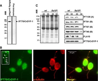
IFT70/CrDYF-1 has a typical localization pattern for IFT proteins, and its entrance into flagella is FLA10 dependent. (A) The wild-type (wt) flagella extract was probed with the affinity-purified α-IFT70/CrDYF-1 antibody with a single band detected. In contrast, preimmune serum does not recognize any specific band. (B) IFT70/CrDYF-1 is localized in the peri-basal body region and flagella. The wt cells were double-labeled with antibodies α-IFT70/CrDYF-1 (green) and α-tubulin (red). The staining with α-tubulin illustrates the position of the two flagella. IFT70/CrDYF-1 is localized primarily in the peri-basal body region as well as in dots along the flagella. The inset shows an enlargement of one of the flagella. (C) The entrance of IFT70/CrDYF-1 into the flagella is FLA10-dependent. Flagellar proteins were extracted from the wt and fla10ts cells after incubation for 50 min at either the permissive temperature (22°C) or the restrictive temperature (32°C), separated on an 8% polyacrylamide gel, transferred to nitrocellulose, and probed with antibodies against IFT70/CrDYF-1 and other IFT complex proteins, as indicated on the right of the Western blots. Equal amount of flagellar proteins were loaded for each sample, as shown by the Coomassie Blue–staining gel in the left panel. The labels A and B represent IFT complexes A and B, respectively.

