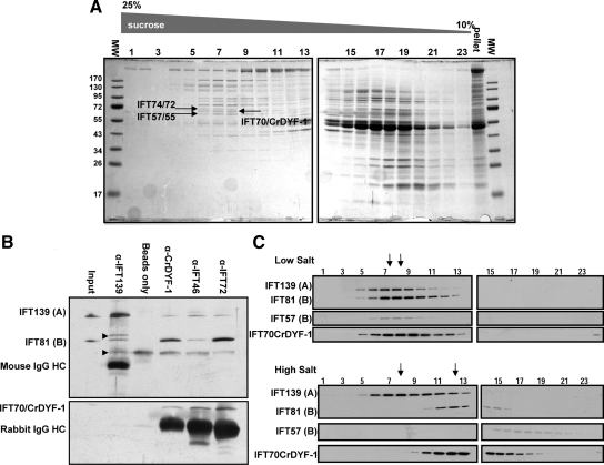
Figure 3.
IFT70/CrDYF-1 is a core component of the IFT particle complex B. (A) IFT70/CrDYF-1 comigrates with other IFT particle subunits at 16S. Flagellar matrix was fractionated through a 12-ml 10–25% sucrose density gradient. The gradient fractions were separated on 10% SDS-PAGE gels and stained with Coomassie Blue. IFT70/CrDYF-1 migrates between IFT72 and IFT57. The lane labeled “pellet” is collected from the bottom of the gradient. (B) IFT70/CrDYF-1 coimmunoprecipitates with other IFT particle complex B proteins. Immunoprecipitates with antibodies against IFT proteins from the flagellar membrane plus matrix were separated on 8% polyacrylamide gels and analyzed by Western blotting. The antibodies used for immunoprecipitation are listed above the Western blots. The antibodies used for Western blotting are indicated on the left. Nonspecific bands are indicated by arrowheads on the left. The band just above IFT81 may come from α-IFT139 antibody, because no such band exists in the starting membrane plus matrix material. The band just below IFT81 apparently comes from protein A beads, as it is present in the beads alone control. (C) Flagellar matrix was treated with or without high salt as described previously (Lucker et al., 2005) and fractionated through a 12-ml 10–25% sucrose density gradient. The sucrose density gradient fractions were separated by 10% SDS-PAGE and analyzed by Western blotting. The arrows mark the peaks of complexes A (left) and B (right).

