Figure 1.
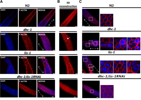
Mutations of dhc-1 and lis-1 disrupt F-actin organization in the pachytene region of the gonad. Phalloidin-rhodamine and Hoechst staining of extruded gonads to visualize F-actin (red) and DNA (blue), respectively. (A) A single optical section of the pachytene germline showing the F-actin structure of the cytoplasmic rachis in wild type (N2), dhc-1(or195ts), lis-1(n3334), and in dhc-1(or195ts); lis-1(RNAi). In wild-type gonads the germline rachis is straight with regularly positioned nuclei in the surrounding cortex, whereas in both mutants the F-actin lining the rachis is ruffled, and many cortical nuclei are displaced into the rachis. (B) 3D reconstruction of F-actin serial optical images to better visualize rachis abnormalities, deformation of the cytoskeleton, and irregularity of actin cages that connect nuclei to the cytoplasmic rachis (arrows). (C) Defects in cortical nuclear localization. Labeled as in A. Images in the leftmost column represent a single optical section near the gonad surface. Areas in white squares are magnified and displayed to the right to illustrate the regular actin network surrounding individual nuclei in wild type and the absence of nuclei or clusters of 2–3 nuclei within an actin ring in both mutants. Distal (D) and proximal (P) orientations of the gonad is indicated on the merged image. Scale bar, 10 μm.

