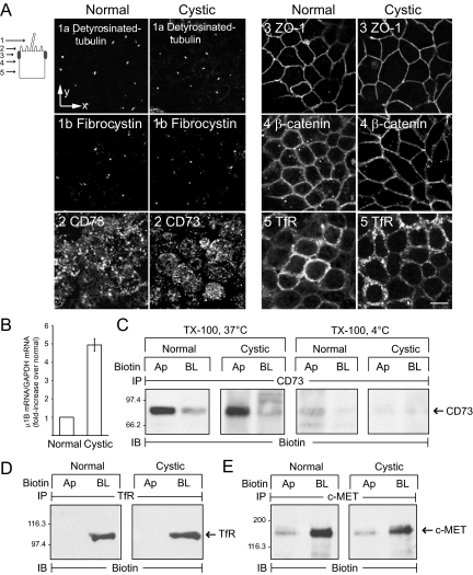
Figure 2.
Epithelial cell markers expressed normally in conditionally immortalized cell lines. (A) Selected vertical x-y confocal optical sections (see schematic) from permeabilized cells stained with antibodies listed in figure. Scale bar, 5 μm. (B) AP-1B μ1B subunit mRNA was measured by qPCR and normalized to an internal GAPDH control. Results for cystic cells (mean ± SEM, n = 4) are presented as fold increase relative to μ1B mRNA expression in normal cells, which was set to 1. (C) Cells were harvested with TX-100 buffer at 37 or 4°C after domain-specific biotinylation. (D and E) Biotinylated cells were harvested with immunoprecipitation buffer at 4°C. (C–E) Cargo-specific immune complexes were immunoblotted for biotin. Ap, apical; BL, basolateral.

