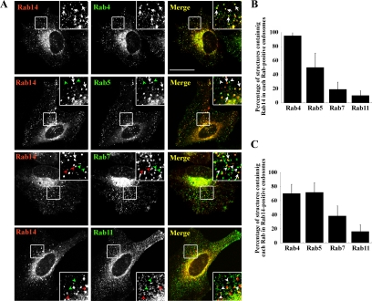
Figure 2.
Overlap of endosomes positive for Rab14 and Rab4. (A) HeLa cells were transiently cotransfected with HA-Rab14 and either EGFP-Rab4, EGFP-Rab5, EGFP-Rab7, or EGFP-Rab11, and processed for immunofluorescence microscopy. Arrows indicate endosomes with overlapping staining for Rab14 and another Rab protein. Green arrowheads, Rab5-, Rab7-, or Rab11-positive endosomes; and red arrowheads, Rab14-positive endosomes. To quantify colocalization, peripheral Rab14-positive endosomes (B) or peripheral endosomes positive for the Rab protein indicated (C) were used as reference. The quantification is based on two independent experiments, using 200–400 endosomes in 6–10 transfected cells. Bar, 20 μm.

