Figure 7.
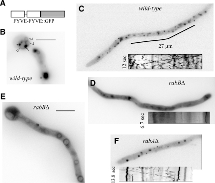
rabBΔ early endosomes are deficient in PI(3)P. (A) The PI(3)P reporter consists of a C-terminal fusion of GFP to two tandem copies of the AnVps27 FYVE domain. (B) (FYVEVps27)2::GFP labels faintly the membrane of basal vacuoles (open arrowheads), which frequently showed closely associated (FYVEVps27)2::GFP punctae (arrows). (C) Tip-proximal region of a wild-type hypha showing localization of (FYVEVps27)2::GFP to small punctae. The kymograph insert (Supplementary Movie 10) illustrates how a proportion of these punctae show the characteristic bidirectional motility of EEs. Relatively immotile ones represent, in all likelihood, late endosomes. (D and E) In rabBΔ cells (FYVEVps27)2::GFP localizes mainly to the cytosol and, in tip-distal regions, also to vacuolar membranes. The few punctae that were discernible against the cytosolic fluorescence haze were immotile (inserted kymograph). (F) rabAΔ cell showing the localization of (FYVEVps27)2::GFP to punctate structures, as in the wild type. Some punctate structures show characteristic EE motility (kymograph).

