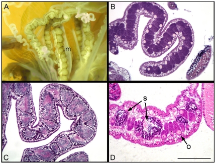
Figure 2. Anatomy and histology of gonads of Aiptasia diaphana.
A) A dissected anemone illustrating the morphology of the gonads along the mesenteries (m) of the polyp. B) Histological section of a female gonad showing well developed oocytes with visible germinal vesicles and nucleoli. C) Histological section of a male gonad showing well developed spermaries. D) Histological section of a hermaphrodite gonad showing well developed oocytes (o) alongside interspersed spermaries (s) at various stages of development. Bar corresponds to 200 µm in B and to 100 µm C and in D.

