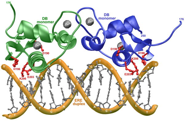
Figure 1.
3D atomic model of the DB domain of human ERα in complex with ERE duplex containing the AGGTCAcagTGACCT consensus sequence. Note that the DB domain binds to DNA as a homodimer. One monomer of the DB domain is shown in green and the other in blue. The Zn2+ divalent ions are depicted as gray spheres and the sidechain moieties of D190, H196, E203 and K206 within the DB monomers are colored red. The DNA backbone is shown in yellow and the bases are colored gray for clarity. The numerals at the termini of DB monomers indicate the boundaries of DB domain within the amino acid sequence of human ERα.

